It’s A Small World After All
In terms of online content, or just shared content in general, videos are taking centre stage and have been challenging the power of photography as of late. To make things a whole lot more interesting, Nikon, a world-renowned and gold-standard brand in the field of optics and imaging, brings us Nikon Small World in Motion Photomicrography Competition for the 11th year in a row.
This year’s lineup of videos features some of the most fascinating and mind-boggling microscopic scenes, submitted by participants from all over the world. If you’re wondering, ‘define microscopic’ or ‘how small is small’, well, picture this…
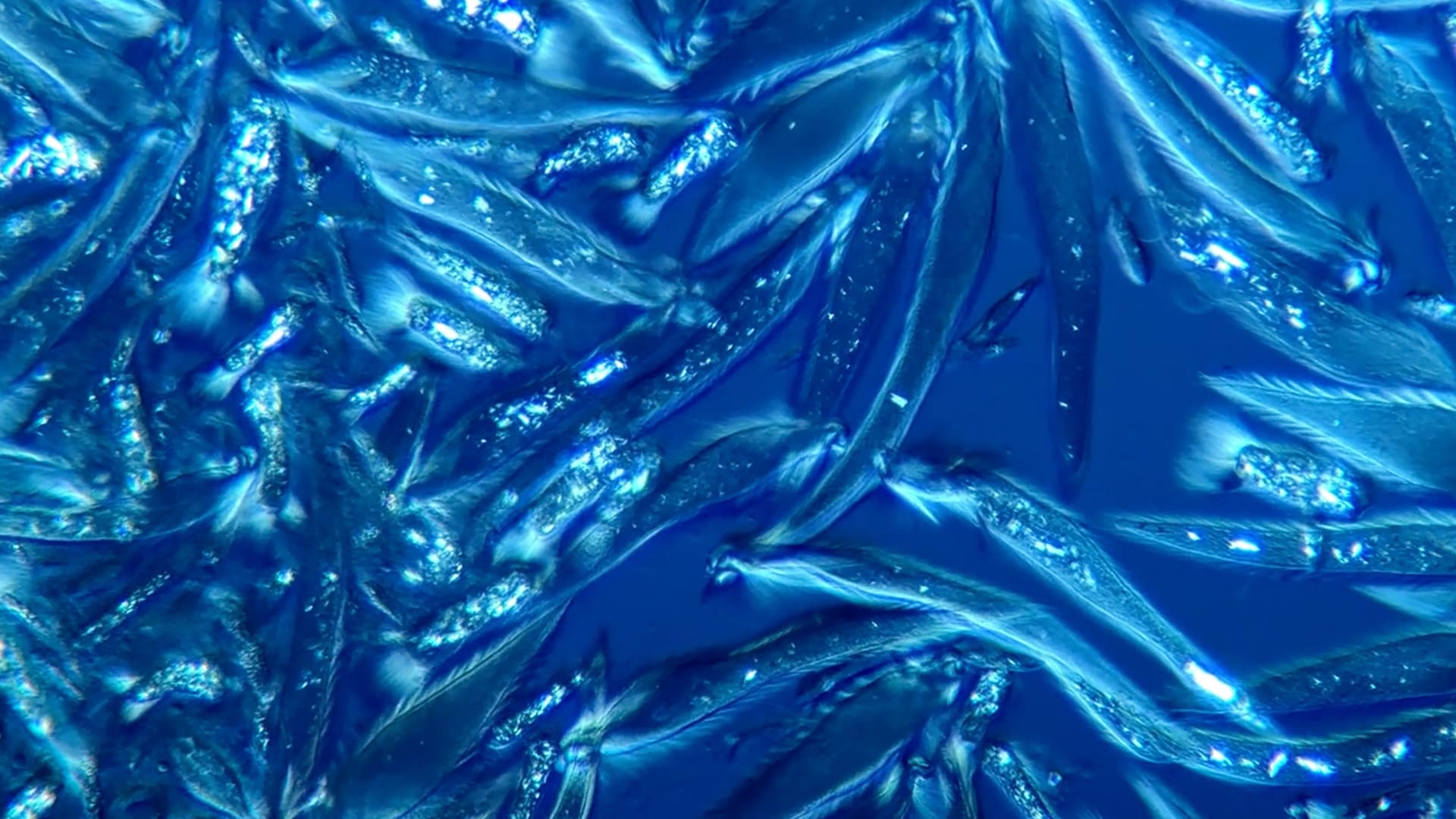
This year’s first-place prize was awarded to Fabian J. Weston for his visually stunning video of live microfauna in the gut of a termite using a research microscope from the 1970s, utilising polarised light. These microorganisms found in termites play the essential role of digesting plant-based cellulose such as wood. Whilst contributing to termite nutrition, their most important role is in carbon cycling. Fabian hopes to visually illustrate the symbiotic relationship between termites and these particular protists, to help audiences better understand the unseen role they play in our natural world.
Fabian adds, “The beautiful thing is that easy access to modern imaging and the internet has allowed those with an interest in microscopy to share their discoveries globally, across all boundaries of culture, language and age. The world is so small, and we can connect easily with anyone across the globe.” Fabian hopes that his winning video will spark greater interest in Protists, as well as inspiring and encouraging more young people to be interested in STEM subjects.
Above: 1st Place Fabian J. Weston, Pennant Hills, New South Wales, Australia | Microfauna in a termite gut | Polarized Light | 10X, 20X & 40X (Objective Lens Magnification)
To see something way beyond what our eyes could see, to share videos that excite, educate, and inspire… to marry creativity and science and produce marvelous things. What a time to be alive?! “We’re living in an amazing time when we have the ability to capture and share high-quality scientific imagery,” said Eric Flem, Communications Manager, Nikon Instruments. “This year’s winning entry highlights the power that microscopy has to connect like-minded individuals, educate others using engaging visuals, and spread scientific knowledge to the general public”
Other notable videos, which are nothing but abstract art perfection in motion, are:
- Dr. Stephanie Hachey and Dr. Christopher Hughes’ 10-day time-lapse of an engineered human micro-tumor forming and metastasising. Vessels (red) support the growing tumor (blue)
- Andrei Savitsky’s video of a water flea (Daphnia pulex) giving birth to cubs shot in 4X (Objective Lens Magnification).
- Danielle Parsons and Alan deHaas’ video of two liquid crystals crystallising on the same microscope slide shot in 20X (Objective Lens Magnification).
- Wojtek Plonka’s ten day time-lapse of moss growth shot in 6X (Objective Lens Magnification)
To view the videos from all winners and honourable mentions, visit nikonsmallworld.com

